FAQs
Frequently asked questions about our solutions
Our wide range of solutions covers the oncology spectrum—from drug discovery to clinical treatment. Browse frequently asked questions about our solutions below.
-
Tumor drug response and
genomic biomarker testing -
3D tumor
models
What is tumor drug-response testing
Drug-response profiling is a treatment selection marker. It determines how an individual cancer patient’s tumor is likely to respond to the various types of chemotherapy by testing the treatment options on that patient’s live cancer cells and measuring cell response. This provides valuable insights that help guide physicians’ treatment decisions, giving you an edge against cancer.
What is genomic testing?
Genomic testing is a group of clinically relevant cancer-related biomarker tests that look at your genetics to predict your cancer’s response to various types of chemotherapy and/or the course your disease is likely to take. The biomarkers included in genomic testing are well-validated and supported by current research and key opinion leaders.
Why have a tumor tested with us?
Your physician is using every advantage available to improve the odds in your fight against cancer. Combining drug-response profiling and genomic profiling of your tumor generates a more comprehensive picture to provide information to help your oncologist individualize your therapy. These tests are designed to reduce your risk of receiving therapies that are ineffective or to identify alternatives when the typical treatments recommended for your type of cancer cannot be tolerated.
How does a tumor get tested?
Tumor tissue is obtained during surgery and your oncologist will send that tissue to our CLIA-certified laboratory using our special collection kit. Two samples of your tumor tissue are obtained, fixed and live. The fixed tumor tissue is used for a panel of genomic biomarker tests. The live tumor tissue is grown in the lab and used to test the drug response of the tumor to a panel of standard-of-care (SOC) drugs. When testing is complete a report is provided back to your clinician with recommended SOC therapies based on the drug response and genomic profiles.
What is the value of having a tumor tested?
The data generated from testing the unique mutations of your tumor and from the drug response profiling, which essentially performs chemotherapy on your tumor outside of the body, provides information to your oncologist to improve the odds in your fight against cancer. In addition, and with your consent, de-identified data generated by testing your tumor is added to our cancer knowledgebase of existing tumor data. Deeper analysis of this data will improve the recommendations we give to clinicians to help patients like you, as well as provide insights to guide research into new cancer therapies.
How much will it cost?
We are committed to providing access to functional precision medicine to as many people as possible. Our panel of biomarker/genomic tests are covered by most insurers/Medicare. We have continued to offer tumor drug-response tests amidst reimbursement uncertainty from both commercial insurers and Medicare. Due to continued clinical demand and interest across the community, we remain committed to meeting the needs of patients. To keep this testing available to patients and their treating physicians, we offer our tumor drug-response profiling at a drastically reduced rate such that no patient will pay more than $615. We also provide a Compassionate Care Program for those patients who need further help with payment. Contact us for more information.
How can I work with 3D tumor models and what are the deliverables?
Contact us by filling out a form at the bottom of this page. We will be in touch to set up a call to discuss the details of your project. We will then provide a budget and timelines based on the study parameters.
For every project you will receive a report summarizing the key findings of the study. Additional deliverables are usually project specific and can include data tables, images, frozen cell pellets and culture supernatants.
I don’t see the system I need. Can I request a custom model?
Absolutely. We love to work on new models and welcome such requests. All custom models are developed by a dedicated team of scientists with expertise in a specific disease or site of interest. To get started, contact us with your model specifications. We will set-up a call to discuss your requirements and model specifications. If necessary, we will put a non-disclosure agreement (NDA) in place to protect any sensitive information. We will then provide you timelines and a budget for the development of your custom model. As a general guideline, a custom model will take 6-9 months to develop and validate with primary cells. You will work with a team of scientists and will receive regular progress reports for each development milestone.
What is 3D culture?
3D culture is a method of culturing cells that promotes formation of 3-dimensional (3D) cellular structures—spheroids, networks, tubules, etc. 3D culture format is preferable to the standard 2D culture, where cells are grown in a monolayer on the surface of cell culture plastic, because it provides a more physiological milieu.
How does our technology differ from other 3D culture platforms?
Our technology is based on 1:1 reconstruction of human tissues comprising of our proprietary organ-specific extracellular matrix (ECM) formulations and disease-specific medium supplements. Our ECM formulations are assembled from natural components. We do not use synthetic hydrogels or plant material; all our matrices have >90% identity with human ECM.
Our models do not simply force cells to clump into 3D structures, they provide a physiologically-relevant milieu where cell-cell and cell-ECM interactions mirror the native architecture of human tissues.
What is our organ-specific ECM?
Each of our organ-specific extracellular matrices is a mixture of ECM components—collagens, laminins, fibronectin, hyaluronic acid, etc.—formulated to mimic the ECM of each human tissue. Once mixed with cells and placed in a well of a cell culture plate it polymerizes to form a semi-solid environment where cells form 3D structures and establish cell-cell and cell-ECM interactions consistent with the architecture of the native tissue.
What is the difference between our various platforms (i.e. r-Bone vs r-Lung)?
Each of our model systems is designed to mimic the organ and the disease of interest. For example, our r-Bone system for multiple myeloma is composed of the bone marrow-specific ECM and multiple myeloma-specific medium supplements. The ECM provides the scaffold mimicking the endosteum and central marrow and the medium supplements provide the multiple myeloma cytokine environment. The same general principle applies to all our model systems.
Are the cells mixed with the ECM or do they grow on top of it?
Both are possible. However, mixing cells with the ECM establishes a more physiologically relevant system. Prior to adding cells, the ECM is a viscous liquid, which polymerizes at room temperature allowing the cells to be suspended within the ECM layer. Growth medium with disease-specific supplements is then overlaid on top of the cell/ECM layer. Over time, cells proliferate and migrate throughout the ECM layer forming 3D structures (i.e. spheroids, tubes, networks).
ls the ECM integrity maintained in presence of proteases produced by the tumor cells?
ECM stays polymerized throughout the course of the culture. However, its stiffness is model-dependent and can become looser when culturing highly aggressive migratory cells.
What is the thickness of your cultures?
The average thickness of the ECM in our models is 800?m-1mm and can be modulated depending on the study goals.
Do therapeutic agents penetrate our ECM after it has polymerized?
Yes. We have tested small molecules, antibodies, antibody-drug conjugates (ADC), and cellular therapies, such as CAR-Ts; in all cases we observe targeted elimination of tumor cells at all depths within the culture.
How long can you maintain primary cells in culture?
Our systems are validated to maintain viability for at least 21 days. However, most models can support primary cells for >30 days. For r-Bone cell cultures we can maintain primary cells in our models for up to 35 days, the maximum duration tested.
What type of therapeutic agents can be tested in reconstructed organ?
Any type of therapeutic agent can be used in reconstructed organ: small molecules, antibodies, antibody-drug conjugates, biologic agents, cell therapeutics (i.e. CAR-T cells).
Why do you wait 3-5 days prior to adding treatment to your culture systems?
We do not add therapeutic compounds to our model systems immediately post setup to avoid artificially skewing the results due to cellular stress and non-physiological cell organization. Culturing cells for 3-5 days gives them a chance to acclimate to the system and to form physiologically relevant cell-cell and cell-ECM interactions that drive the environmentally-mediated drug resistance (EMDR). Setting-up proper tissue architecture is crucial for the accurate prediction of drug response.
What should be the sample viability to obtain successful growth in the culture?
For successful cultures, the cells must retain >70% viability after thaw.
What is r-Bone?
Reconstructed Bone (r-Bone) is a 3D cell culture model system designed to reconstruct the bone marrow tissue for long-term culture of primary bone marrow cells. The first component of the r-Bone model is the ECM that provides the semi-solid scaffold reconstructing two distinct niches within the human bone: (1) the the endosteum, an interphase between the solid bone and the bone marrow, and (2) the central marrow. The second component is the medium supplement is formulated to mimic the disease-specific circulatory environment.
What indications have you tested in r-Bone?
The r-Bone system has been tested with primary bone marrow cells from patients with AML, multiple myeloma, MGUS, plasma cell leukemia, amyloidosis, as well as non-malignant bone marrow from healthy donors.
Can you culture both fresh and cryopreserved bone marrow samples in r-Bone?
Yes, both fresh and viably cryopreserved bone marrow samples can be cultured in r-Bone. Fresh samples are first run through a Ficoll gradient to isolate bone marrow mononuclear cells (BMMCs), which are then mixed with the r-Bone ECM and plated. Cryopreserved BMMCs are directly mixed with the r-Bone ECM without additional processing. For successful cultures, the cells must retain >70% viability after thaw.
Does r-Bone support both hematopoietic and stromal compartments of the bone marrow?
Yes. Lymphoid, myeloid, and stromal populations present in the patient sample prior to culture are present after 21 days of culturing in r-Bone.
Do malignant plasma cells from the BMMCs of multiple myeloma patients expand in r-Bone?
Yes. The primary malignant multiple myeloma plasma cells proliferate in r-Bone. The extent of the expansion is patient dependent, but we routinely observe 2-10-fold expansion of the malignant clone.
Can I follow proliferating cells in r-Bone?
Yes, BMMCs can be labeled with CFSE prior to plating in the r-Bone ECM. Proliferation can then be observed by microscopy, flow cytometry, or fluorescence measurement on a plate reader.
Do you enrich for multiple myeloma plasma cells before culturing in r-Bone?
No. To retain the heterogeneity of the ex vivo samples, we culture the mononuclear cells obtained from the bone marrow aspirates through Ficoll gradient.
Have you tested standard of care drugs against multiple myeloma in r-Bone?
Yes. Some of the drugs that have been tested in the r-Bone include: bortezomib, carfilzomib, lenalidomide, pomalidomide, daratumumab, dexamethasone and various combinations, such as RVD and CPD.
What readout is compatible with our technology?
Our technology is compatible with any standard readout techniques, including but not limited to, flow cytometry, microscopy, plate reader-based cell assays (CellTiter Glo, MTS, etc.), nucleic acid analysis (RNAseq, qPCR, arrays, etc.), proteomics, and in vivo studies.
Cells can be isolated from the organ-specific ECM using our proprietary non-enzymatic isolation solution that is compatible with flow cytometry and any other readout strategies that require intact surface receptors. Moreover, cells remain viable after isolation and can be subsequently used in both in vitro and in vivo studies.
What is the detection limit in our models?
The dynamic range of CellTiter Glo assay in our models is 10-80,000 cells per well in a 96-well plate.
Is it possible to image through the culture?
Yes, in-matrix imaging is possible. Our 3D tumor models systems have been tested with brightfield, fluorescence, confocal and high content imaging methods.
What is the cell viability and yield post isolation?
We routinely obtain cells with >70% viability post isolation from untreated cultures. The yield is model dependent since different numbers of cells are plated per well depending on the tissue being reconstructed. For example, in rBone the yield from a single well of a 96-well plate is approximately 100,000 cells, but from r-Lung, it is ~20,000 as the plating densities and proliferation properties of the cells differ significantly between these models.
Do you get enough material for gene expression analysis?
Yes. The number of wells required to obtain enough material will depend on the model type and cell plating density.
Let’s discover and develop together.
Complete the form and we’ll be in touch to discuss customized solutions for your needs.
News & resources
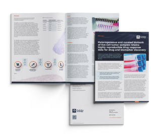
White paper validates highly reproducible drug response data. Results enable AI platform to model and predict patient outcomes on historical samples Leveraging more than 20...
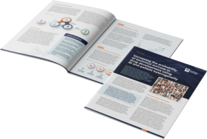
A recent white paper released by Predictive Oncology’s highlights the challenge of late-stage clinical trial failures and the company’s ability to better navigate those obstacles...
Company expands AI/ML driven capabilities to pursue novel biomarker discovery to predict patient outcomes and drug response in oncology. Read our white paper below that...
